Have you ever wondered how small objects you can see? From a grain of sugar in coffee, to cells in a strand of hair or cheek, some can only be examined with the naked eye. If these items are already hard to examine, what about smaller parts of organisms and other things that are barely visible? What do we do? That's what microscopes are for.
The microscope was first developed in 1590 by Janssen and son in the Netherlands. It's a tool for looking at smaller objects that the human eye can't see. There are many types of microscopes. The most common is an optical microscope, which uses a light image of a sample.

Other major types of microscopes are electron microscopes, ultramicroscopes, and various types of scanning probe microscopes. Microscopy is the scientific field of study used for the study of small structures and objects by means of a microscope. Let's take a look at the role of microscopes and their classification.
(1) The structure and function of the microscope
1, eyepiece: zoom object image, 5X, 10X, 40X and so on.
2. Lens tube: connect eyepiece and objective lens.
3, converter: the joint of the objective lens, used to convert the objective lens.
4, objective lens: magnify the object image, there are generally three, magnification is generally 4X, 10X, 40X.
5. Loading table: slide specimens are placed with a tablet clamp.
6, light hole: through the light.
7, sunshade: adjust the light intensity.
8. Tablet holder: Fix the glass specimen.
9. Coarse quasi focusing screw: adjust the focal length and use it when viewing with low power lens. High power lens cannot be used.
10, reflector: light into the lens tube (now the microscope generally do not have this structure, are power supply, more convenient).
11. Fine focusing screw: adjust the focal length and use the high power lens for observation.
12. Mirror arm: it connects the mirror base and the mirror body, and holds the mirror body. The right hand holds the mirror arm when using.
13. Mirror column: support the mirror body.

(2) How does a microscope work?
Basic microscopes used in various structures today use a series of lenses to collect, reflect, and focus light into the sample that is being examined. A microscope will not work without light. Such microscopes are commonly used in research centers, schools and hospitals.
Using different microscope lenses can increase magnification without changing the quality of the resulting image. In addition to magnifying the lens, it is also important to recognize the microscope's field of view to accurately measure specimen size.
Also, most microscopes have a binocular, which consists of two lenses and a prism for separating the image from the two eyepieces you will be looking into.
At the other end of the microscope is an objective lens, which collects and concentrates light into the sample. These objects have different strengths and can be used one at a time by adjusting the rotating frame.

An instrument called an eyepiece magnifies objects by changing the wavelength of light used in the instrument. There are many types of eyepieces, each capable of performing different tasks.
Common eyepieces are those that use gas displacement technology to provide light. The next common eyepiece is the gas correction model. The third common eyepiece is the photocell model.
Other types of eyepieces also exist and are used according to experimental needs. By understanding how microscopes work, researchers will be able to use these eyepieces in experiments, providing a better way to study nature and how it works.
Microscopes are usually powered by batteries or mechanical devices to view objects up to 10 times smaller than their original size. If the specimen under the microscope is not handled properly or improperly, the image can be distorted and give misleading results.
Therefore, using the right type of microscope and handling it correctly is important to observe the object you have chosen.

(3) Microscopic classification
Microscopes can be divided into polarizing microscope, optical microscope, electron microscope and digital microscope.
Optical microscope: usually consists of an optical part, an illumination part and a mechanical part. No doubt the optical part is the most critical, it consists of eyepiece and objective lens.
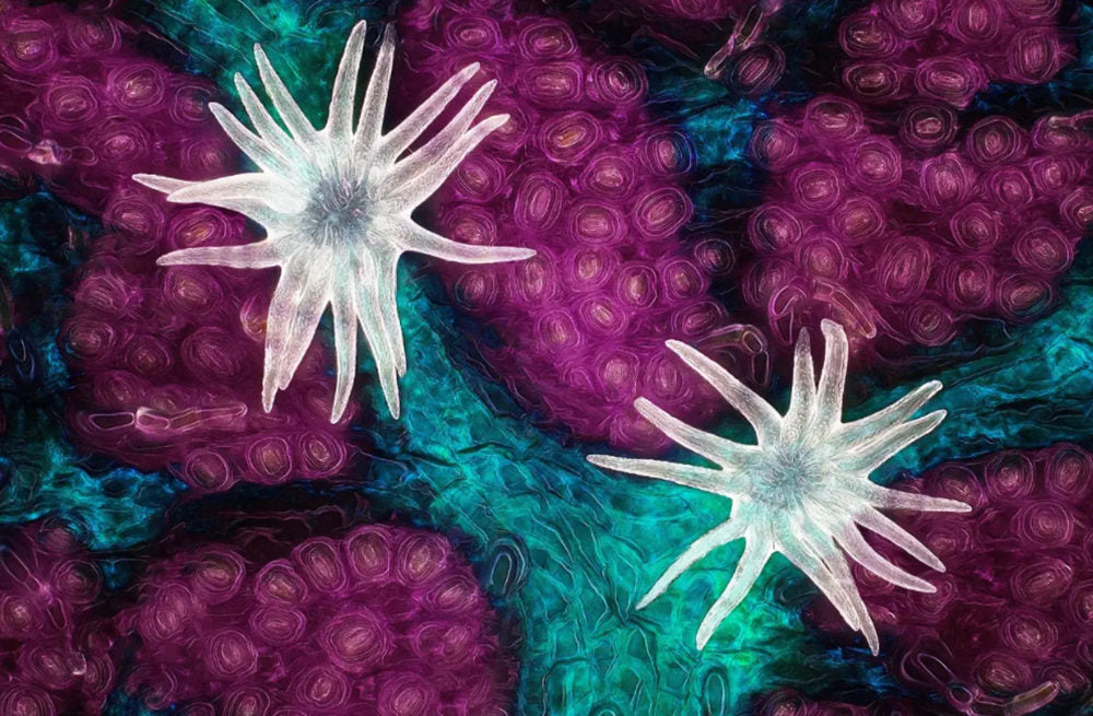
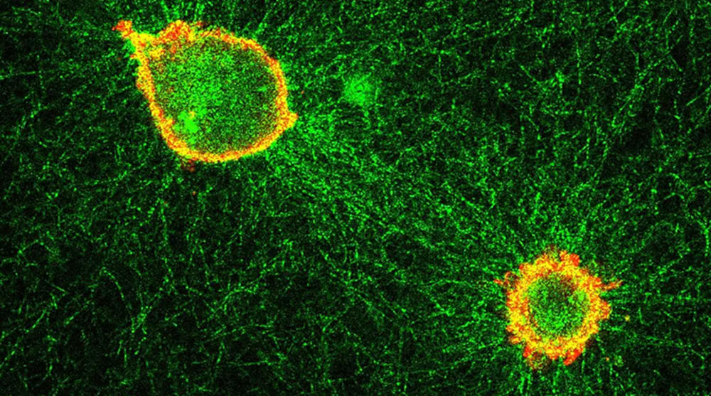
There are many kinds of optical microscope, including bright field microscope (ordinary optical microscope), dark field microscope, fluorescence microscope, phase contrast microscope, confocal laser scanning microscope, polarized light microscope, differential interference difference microscope, inverted microscope.
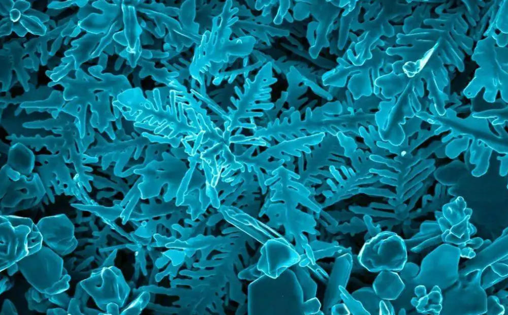
Electron microscope: electron microscope has much higher magnification and resolution power than optical microscope. It uses electron stream as a new light source to image objects. Types of electron microscopes include transmission electron microscopes, X-ray microscopes and scanning electron microscopes. Often used in biology, medicine and small particle observation.

Polarized light microscope: where there is birefringence of the material, under the polarized light microscope can be distinguished clearly, of course, these substances can also be observed using staining method, but some are not available, and must use the polarized light microscope. Polarizing microscope is mainly used to observe liquid crystal, fiber and other objects with single refraction or birefraction.
Digital microscope: digital microscope is a high-tech product which combines the excellent optical microscope technology, advanced photoelectric conversion technology and LCD screen technology perfectly. Thus, we can study the microscopic field from the traditional ordinary eyes to be observed through the display, thus improving the work efficiency.
By the end of this tutorial you may have a basic understanding of microscopes, whether you are ready to buy a microscope or have already bought one and are ready to try it out. Stay tuned for more microscopes on our blog.


Share:
Composition Tips For Architecture Photography
Professional light microscope type